Lab-grown ‘mini brains’ produce electrical patterns that resemble those of premature babies
Table of Contents

The advancement could help scientists to study early brain development. Research in this area has been slow, partly because it is difficult to obtain fetal-tissue samples for analysis and nearly impossible to examine a fetus in utero. Many researchers are excited about the promise of these ‘organoids’, which, when grown as 3D cultures, can develop some of the complex structures seen in brains.
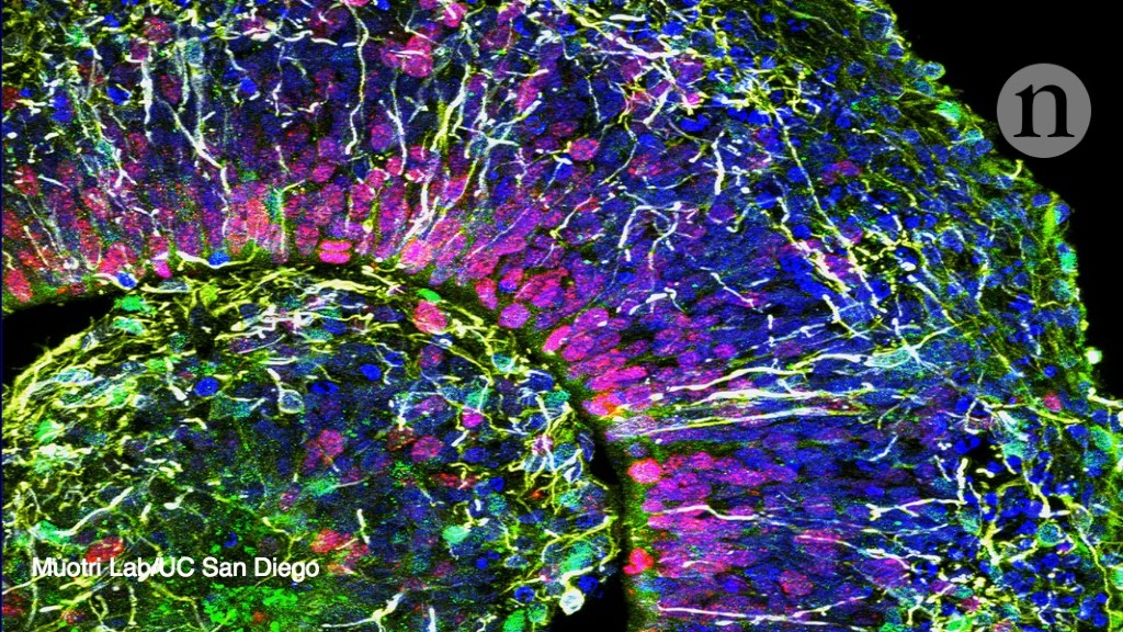
But the technology also raises questions about the ethics of creating miniature organs that could develop consciousness. A team of researchers led by neuroscientist Alysson Muotri of the University of California, San Diego, coaxed human stem cells to form tissue from the cortex — a brain region that controls cognition and interprets sensory information. They grew hundreds of brain organoids in culture for 10 months, and tested individual cells to confirm that they expressed the same collection of genes seen in typical developing human brains1.
The group presented the work at the Society for Neuroscience meeting in San Diego this month. Muotri and his colleagues continuously recorded electrical patterns, or electroencephalogram (EEG) activity, across the surface of the mini brains. By six months, the organoids were firing at a higher rate than other brain organoids previously created, which surprised the team.
The EEG patterns were also unexpected. In mature brains, neurons form synchronized networks that fire with predictable rhythms. But the organoids displayed irregular EEG patterns that resembled the chaotic bursts of synchronized electrical activity seen in developing brains.
When the researchers compared these rhythms to the EEGs of premature babies, they found that the organoids’ patterns mimicked those of infants born at 25–39 weeks post-conception.
Source: nature.com

