Human blood cells transformed into functional neurons

Human blood cells transformed into functional neurons

  • June 6, 2018
Table of Contents

Human blood cells transformed into functional neurons

Human immune cells in blood can be converted directly into functional neurons in the laboratory in about three weeks with the addition of just four proteins, researchers at the Stanford University School of Medicine have found. The conversion occurs with relatively high efficiency — generating as many as 50,000 neurons from 1 milliliter of blood — and it can be achieved with fresh or previously frozen and stored blood samples, which vastly enhances opportunities for the study of neurological disorders such as schizophrenia and autism. A paper describing the findings was published online June 4 in the Proceedings of the National Academy of Sciences.

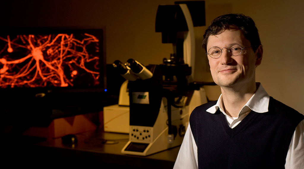
Wernig is the senior author. Former postdoctoral scholar Koji Tanabe, PhD, and graduate student Cheen Ang are the lead authors.

Source: stanford.edu

Share :
comments powered by Disqus

Related Posts

Leading UK scientist reveals likely cause of childhood leukaemia

Leading UK scientist reveals likely cause of childhood leukaemia

Professor Mel Greaves from The Institute of Cancer Research, London, assessed the most comprehensive body of evidence ever collected on acute lymphoblastic leukaemia (ALL) – the most common type of childhood cancer. His research concludes that the disease is caused through a two-step process of genetic mutation and exposure to infection that means it may be preventable with treatments to stimulate or ‘prime’ the immune system in infancy. The first step involves a genetic mutation that occurs before birth in the foetus and predisposes children to leukaemia – but only 1 per cent of children born with this genetic change go on to develop the disease.

Read More
Mice with 3D-Printed Ovaries Successfully Give Birth

Mice with 3D-Printed Ovaries Successfully Give Birth

AsKatherine Kornei at Science reports,the researchers used a 3D printer to build the scaffolding of the organs, weaving layers of gelatin to createtiny (15 x 15 millimeter) ovaries on glass slides. They then tested the scaffolds by embedding a follicle—the tiny sacs composed of hormone-secreting cells that containthe maturing eggs. This test suggested that the tightest weave supported the highest survival rates, reports Kornei.

Read More