First ever color X-ray on a human

First ever color X-ray on a human

  • July 13, 2018
Table of Contents

First ever color X-ray on a human

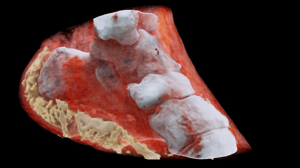
New Zealand scientists have performed the first-ever 3-D, colour X-ray on a human, using a technique that promises to improve the field of medical diagnostics, said Europe’s CERN physics lab which contributed imaging technology. The new device, based on the traditional black-and-white X-ray, incorporates The CERN technology, dubbed Medipix, works like a camera detecting and counting individual sub-atomic particles as they collide with pixels while its shutter is open.

This allows for high-resolution, high-contrast pictures. The machine’s ‘small pixels and accurate energy resolution meant that this new imaging tool is able to get images that no other imaging tool can achieve,’ said developer Phil Butler of the University of Canterbury. According to the CERN, the images very clearly show the difference between bone, muscle and cartilage, but also the position and size of cancerous tumours, for example.

The technology is being commercialised by New Zealand company MARS Bioimaging, linked to the universities of Otago and Canterbury which helped develop it.

Source: yahoo.com

Share :
comments powered by Disqus

Related Posts

How gut microbes are joining the fight against cancer

How gut microbes are joining the fight against cancer

Cancer has been a late bloomer in the microbiome revolution that has surged through biomedicine. Over the past few decades, scientists have linked the gut’s composition of microbes to dozens of seemingly unrelated conditions — from depression to obesity. Cancer has some provocative connections as well: inflammation is a contributing factor to some tumours and a few types of cancer have infectious origins.

Read More
Our cancer preventing genes revealed

Our cancer preventing genes revealed

In our bodies, we all have genes working hard to prevent cancer. If they don’t do their job properly, rogue cells can mutate and develop into the life-threatening disease. The malfunction of one so-called “super tumour suppressor gene” known as p53 causes at least half of all cancers.

Read More
Leading UK scientist reveals likely cause of childhood leukaemia

Leading UK scientist reveals likely cause of childhood leukaemia

Professor Mel Greaves from The Institute of Cancer Research, London, assessed the most comprehensive body of evidence ever collected on acute lymphoblastic leukaemia (ALL) – the most common type of childhood cancer. His research concludes that the disease is caused through a two-step process of genetic mutation and exposure to infection that means it may be preventable with treatments to stimulate or ‘prime’ the immune system in infancy. The first step involves a genetic mutation that occurs before birth in the foetus and predisposes children to leukaemia – but only 1 per cent of children born with this genetic change go on to develop the disease.

Read More